Many women recognise the pattern. A routine procedure takes longer than expected. It’s more uncomfortable than promised. The doctor reassures them that this sometimes happens, or suggests anxiety or muscle tension might be playing a role. But often the explanation is simpler – and anatomical.
This mismatch between bodies and procedures isn’t related to rare conditions or specialist care. It reflects a recurring problem in everyday medicine. Many routine procedures were designed around male anatomy, and they don’t always work the same way on female bodies.
Take colonoscopy. It’s one of the most common investigations used to diagnose bowel disease and screen for cancer. Yet women are more likely than men to experience discomfort, require repositioning, or have an incomplete examination on the first attempt.
The reason lies in normal anatomy. On average, women have a longer and more mobile colon, particularly in the sigmoid segment that loops through the pelvis.
The female pelvis itself is broader and shallower, creating sharper angles as the bowel curves downward. These features make the scope more likely to bend and loop inside the bowel, slowing its progress and pulling on surrounding tissue – a major source of pain.
This isn’t abnormal anatomy. It’s normal anatomy that standard techniques don’t always take into account.
Urinary catheterisation is another routine procedure where anatomy matters. Although the urethra performs the same function in men and women, its length, course and anatomical context differ in ways that matter clinically.
In males, the urethra is long – around 18-22cm – and is usually described in three parts: the prostatic urethra, which is wide and fixed as it passes through the prostate; the membranous urethra, the narrowest segment as it crosses the pelvic floor; and the spongy (penile) urethra, which runs in a predictable course to a clearly identifiable external opening at the tip of the penis. Despite its length, the male urethra follows a stable path and ends at a prominent external landmark.
The female urethra is much shorter, usually about 3-4cm long, but lies within a more variable anatomical environment. From the bladder neck, it passes through the bladder wall and pelvic floor, before opening into the vulval vestibule at a meatus (the external opening of the urethra) closely related to the anterior vaginal wall.
Its position varies between individuals and across the life course, influenced by pelvic floor tone and hormonal status. In practice, this can make catheter insertion technically more difficult, increasing the likelihood of repeated attempts and discomfort – particularly in older women or those with atrophic tissue (thin, delicate tissue).
Lumbar puncture and spinal procedures show similar issues. Women tend to have a greater lumbar curve and different pelvic tilt, altering the angle at which a needle must pass between vertebrae. Mild spinal curvature is also more common in women. The procedure itself doesn’t change, but the geometry does, increasing the likelihood of multiple attempts and prolonged discomfort.
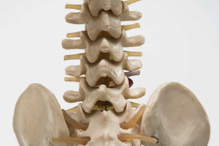
Teeradej/Shutterstock.com
Even airway management, a cornerstone of anaesthesia and emergency medicine, reflects the same mismatch. Female airways are, on average, shorter and narrower. When equipment sizing and technique is based on a “standard” airway, women are more likely to experience sore throat and hoarseness afterward – effects often dismissed as minor, but rooted in anatomy rather than sensitivity.
Even something as commonplace as peripheral venous cannulation, the insertion of a small tube into a vein to deliver fluids, medications, or to take blood, reflects this mismatch. Women’s superficial veins are often smaller, less prominent and more mobile in soft tissue, making standard cannulation techniques more likely to result in repeated attempts, bruising and pain.
Design for variation, not exception
Doctors know bodies vary. In practice, many already adapt – choosing different patient positions, smaller instruments or altered techniques. But these adjustments are informal, inconsistently taught and rarely explained to patients.
Instead, difficulty is often bundled into vague categories: anxiety, tension, low pain tolerance or “one of those things”. The result is that women experience real, anatomy-driven discomfort without being told why, and may internalise it as a personal failing.
This matters. When discomfort is normalised or minimised, patients are less likely to return for screening, more likely to delay care, and more likely to mistrust reassurance that future procedures will be different.
None of this requires radical innovation. It requires naming the issue accurately. When procedures are taught and designed around a single reference body, predictable anatomical variation becomes an obstacle rather than a design feature.
Acknowledging that bodies differ – in length, curvature, mobility and spatial relationships – allows doctors to plan, explain and adapt more effectively.
Crucially, it also shifts the narrative. Instead of “this shouldn’t hurt”, the message becomes: “your anatomy means this procedure can be more challenging, and we’ll adjust it accordingly”.
![]()
Michelle Spear does not work for, consult, own shares in or receive funding from any company or organisation that would benefit from this article, and has disclosed no relevant affiliations beyond their academic appointment.